MSc Research Project – SynthBA & Brain-Age Prediction
“Contrast and Resolution Agnostic Brain-Age Prediction for Predicting Neurodegeneration“

In my Master’s thesis project, I investigated how single- and multi-modal MRI data can be used to predict the biological brain age with the use of a contrast-agnostic deep learning framework, SynthBA. Using the large-scale ADNI database, I examined how imaging modality, resolution, and diagnosis influence the prediction performance and reproducibility.
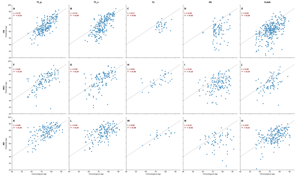
I explored whether SynthBA could provide reliable, clinically meaningful brain-age estimates across heterogeneous MRI scans, including T1w, T2w, PD, and FLAIR images at resolutions of 1mm and 2mm. My work evaluated prediction error (MAE, R2) and group differences in cognitively normal (CN), mild cognitively impaired (MCI), and Alzheimer’s disease (AD) participants, as well as the brain-PAD (brain-predicted age difference), with its correlation with cognitive decline using ADAS-Cog13 scores.
This research highlights the potential future of contrast-agnostic brain-age models for real-world applicability and identifies key limitations when generalising across modalities.
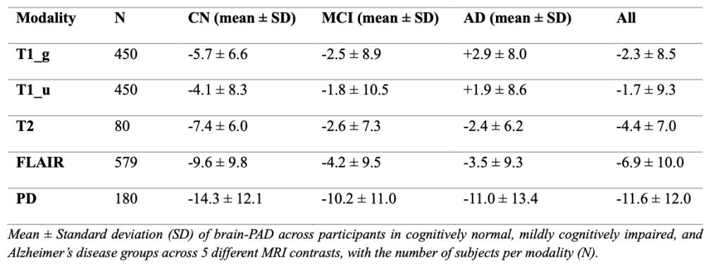
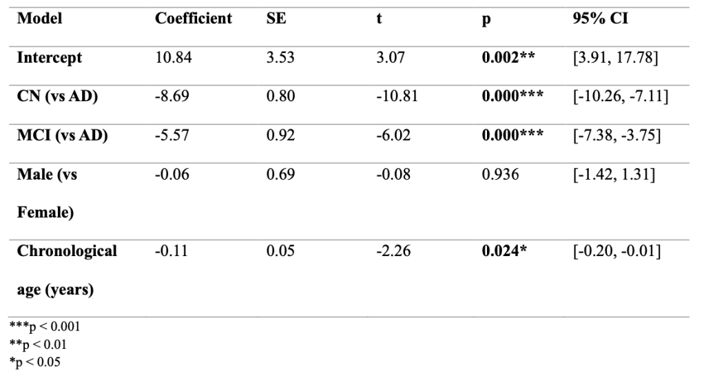
I found that the predicted brain age generally exceeded the chronological age in the MCI and AD groups, resulting in a weaker correlation and a more positive brain-PAD, consistent with accelerated ageing (Table 1). To estimate the impact of diagnosis on brain-PAD derived from T1w-MRIs in model g, a linear regression was investigated (as shown in Table 2). CN subjects had a statistically significant difference with a lower brain-PAD than AD subjects by approximately 8 years (β = -8.69, p < 0.001).
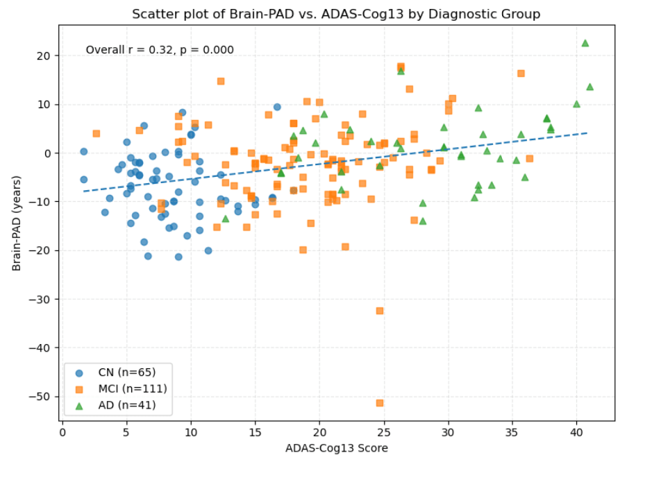
Correlation between Brain-PAD and Cognitive Impairment
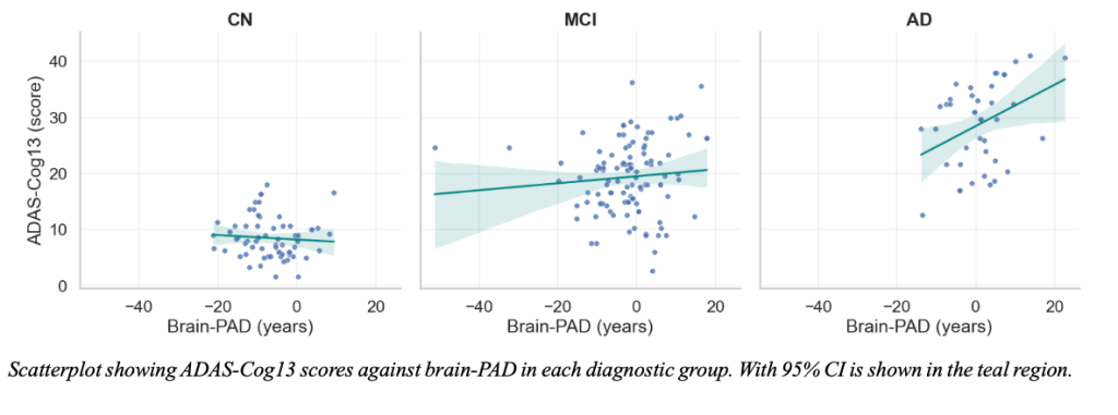
As assessed with ADAS-Cog13 scores, there was a significant positive Pearson’s r correlation (r = 0.32, p < 0.001), indicating that higher brain-PAD was associated with worse cognitive function (higher ADAS-Cog13 scores) (Fig. 2). When stratified by diagnostic group, the relationship was particularly driven by AD participants which demonstrated a moderate positive association between brain-PAD and ADAS-Cog13, with no significant correlations found in CN and MCI participants (Fig. 3).
Overall, this research demonstrates SynthBA’s ability to serve as a reliable brain aging biomarker over other brain age models with the ability to perform robustly across multiple MRI modalities and resolutions with minimal preprocessing needed. While most approaches are restricted to T1w MRIs and extensive pipelines, SynthBA was able to generate accurate predictions under variable imaging conditions, which is more reflective of real-world clinical data. Furthermore, the investigation of the ensemble approach in compensating for weaker inputs emphasises the robustness of the model despite not exceeding the best-performing single modality. These findings highlight SynthBA’s potential to adapt to heterogeneous imaging protocols.
Future research:
- Tracking brain age over time to monitor brain health
- Can determine whether this potential brain age biomarker can predict cognitive decline using brain-PAD values
- Further analyses into ensemble approach should be investigated with a more balanced sample size across modalities and diagnostic groups to improve generalisability
- Further validation from routine clinical scans to be tested in SynthBA to test for robustness under extensive variability in healthcare settings
